Switching immune cells on is a critical step in combating infection. In the case of T cells, which are key players in immunity, this happens when dormant T cells circulating in the blood encounter a suspicious agent on the surface of specialized antigen-presenting cells (APCs). APCs detect and pick up foreign agents they encounter as part of routine immune surveillance.
In order for T cells to get a good read on foreign molecules, they must establish a close communication interface with APCs. This interface, known as an immunological synapse, is vital to T-cell activation. Researchers have been doggedly pursuing a greater understanding of what happens during synapse formation and how these events shape immune responses against everything from the common cold to novel infections to cancer.
Now, thanks to a new $1.98M grant from the National Institutes of Health National Institute of Allergy and Infectious Diseases (NIAID), scientists at the Lewis Katz School of Medicine at Temple University (LKSOM) are poised to make major headway into the study of immunological synapse formation.
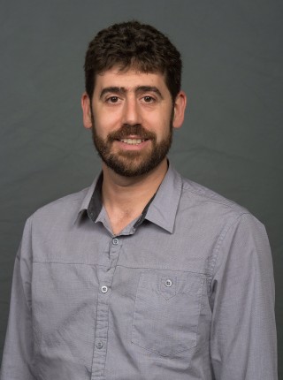
“T-cell activation is vital for immunity, for fighting off infectious diseases,” said Jonathan Soboloff, PhD, Professor of Medical Genetics and Molecular Biochemistry at the Fels Institute for Cancer Research and Molecular Biology at LKSOM, and senior investigator on the new grant. “We're living through a pandemic right now, and COVID-19 is showing us why it is so important to develop a deeper knowledge of the mechanisms underlying T-cell activation.”
A major goal of the research funded by the new NIAID award is to identify the role in immunological synapse formation of a molecule known as STIM1. STIM1 is a calcium sensor located within the endoplasmic reticulum, a continuous membrane system in the cell that functions in part in protein folding and transport. In previously published work within the field, STIM1 was shown to move to the side of the T cell where the immunological synapse forms. Dr. Soboloff and colleagues have found that STIM1 localization to the immunological synapse raises local calcium levels, leading to the loading of calcium into mitochondria – the energy-supplying powerhouses of cells.
These observations suggest that STIM1 supports mitochondrial function during T-cell activation. “Using novel STIM1 mutants, we will determine what makes STIM1 move to the immunological synapse during T cell activation,” Dr. Soboloff said. “This will also allow us to characterize its impact on cellular metabolism to better define how STIM1 and calcium regulation impact T-cell activation.”
Under the new grant, Dr. Soboloff also plans to investigate the role of proteins known as septins in immunological synapse formation and the activation of immune cells. Septins provide a sort of scaffolding inside cells that influences protein movement to specific locations and that creates barriers to prevent proteins from leaving cellular compartments. While septins are known to contribute to STIM1 function in some contexts, there are no publications regarding their involvement in protein localization during the formation of the immunological synapse.
“Ultimately, through experiments in cell lines and mouse models, we want to identify the underlying molecular mechanisms driving STIM1 translocation and determine the physiological relevance of this process,” Dr Soboloff explained. “In addition to extending our understanding of STIM1 in the context of T-cell activation, new knowledge of how these mechanisms are applied could lead to novel insight into the significance of STIM1 regulation in cellular metabolism and its implications more generally in cell biology.”
The NIAID award provides support for Dr. Soboloff's research into STIM1 and T-cell activation through March 2025. Co-investigators on the award include Yi Zhang, MD, PhD, Professor of Microbiology and Immunology at the Fels Institute for Cancer Research and Molecular Biology, and John Elrod, PhD, Associate Professor of Pharmacology and Associate Professor at the Center for Translational Medicine and the Alzheimer's Center at Temple.
Bottom image description: During T-cell activation, many cellular components reorient themselves towards a specialized area of the cell, the immunological synapse. These images taken by TIRF microscopy show the bottom of the cell only. In resting cells, STIM1 is localized throughout the cell (upper images), but when T cells are activated, it relocalizes to a confined area. The discovery of a mutation that blocks this relocalization during T-cell activation is key to understanding the contribution of STIM1 localization at the immunological synapse to T-cell activation.
