The immune system is like a carefully regulated machine, complete with its own built-in “brakes” that prevent it from overreacting and causing excess inflammation in otherwise healthy tissues. This preventative safety net , however, is highly vulnerable, particularly in cancer, where tumor cells step on the brakes constantly, because doing so allows the tumor cells to escape immune detection.
Several molecules that act as natural brakes on immune activity have been discovered, which has opened the door to immunotherapy – a potentially highly effective way of leveraging the immune system to attack cancer cells. For immunotherapy to reach its full potential in human patients, however, more must be learned about factors driving cancer immunity.
Now, researchers at the Lewis Katz School of Medicine at Temple University (LKSOM) and Fox Chase Cancer Center show for the first time that a molecule called EGR4 –known mainly for its role in male fertility – serves as a critical brake on immune activation. The new study, published online March 25 in the journal EMBO Reports, shows that taking EGR4 away – effectively releasing the brake – promotes the activation of so-called killer T cells, which infiltrate and attack tumors and thereby boost anticancer immunity.
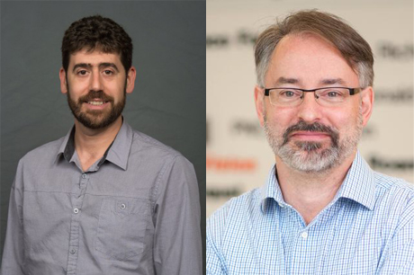
“Other early growth response proteins, or EGRs, are important to T cell activity, but whether EGR4 also has a role in immunity has been largely overlooked due to early challenges in its detection,” explained Jonathan Soboloff, PhD (pictured above, left), Professor of Medical Genetics and Molecular Biochemistry at the Fels Institute for Cancer Research and Molecular Biology at LKSOM. “Our study reveals a new side to the importance of EGR4.”
After early findings in T cell lines led Dr. Soboloff's team (Robert Hooper, PhD, and Elsie Samakai, PhD) to suspect that EGR4 had a role in T cell biology, they obtained EGR4 knockout mice from Dr. Warren Tourtellotte (Cedars Sinai Medical Center; Los Angeles, CA). They further established that EGR4 was expressed in WT T cells upon stimulation and that loss of EGR4 led to a T cell phenotype.
“We know from our previous work that T cells control calcium signaling and that when intracellular calcium levels are elevated, calcium signaling can drive T cell activation,” Dr. Soboloff said.
To better define the immunological role of EGR4, Dr. Soboloff collaborated with Dietmar J. Kappes, PhD (pictured above, right), Professor, and Jayati Basu, PhD, Research Associate of the Blood Cell Development and Function Program at Fox Chase Cancer Center, due to their expertise in this field.
Remarkably, this investigation revealed that loss of EGR4 leads to dramatic increases in calcium signals in T cells, leading to the specific expansion of T helper type 1 (Th1) cell populations. Th1 cells represent a key cell population that act as activators of cytotoxic, or killer, T cells, which are responsible for destroying tumor cells and virus-infected cells. The team next recruited Mr. Scott Gross (Soboloff Lab), who worked together with Drs. Kappes and Basu to investigate the functional importance of EGR4 in cancer immunity by utilizing an adoptive mouse model of melanoma in which some host animals lacked EGR4 expression. Compared to mice with typical EGR4 levels, EGR4 knockout animals showed strongly enhanced anticancer immunity. In particular, EGR4 knockout mice had reduced lung tumor burden and fewer metastases than mice with normal EGR4 expression.
In future work, the team plans to further explore strategies for EGR4 targeting. The development of an agent to target EGR4 specifically may be difficult, due to the diverse actions of EGR pathways. “But eliminating EGR4 specifically from a patient's T cells, and then putting those cells back into the patient, may be a viable immunotherapeutic approach,” Dr. Kappes said.
This work was accomplished by a strong team effort where Dr Soboloff’s team (Drs. Robert Hooper, Christina K. Go, Elsie Samakai, Scott Gross and Bryant Schultz) with great expertise in calcium signaling, were complemented by Dr. Kappes and Dr. Basu’s extensive expertise in T cell development, function, and cancer immunology, as well as Dr. Raza Zaidi’s expertise in melanoma pathology. Ultimately, this investigation provided an optimal example of scientific collaboration amongst senior and junior scientists located at different Institutes within the Temple University umbrella. Other investigators who contributed to this new study include Jonathan Ladner, Blood Cell Development and Function Program, Fox Chase Cancer Center; and Yuanyuan Tian, Bo Zhou, Shan He, and Yi Zhang, Fels Institute for Cancer Research and Molecular Biology and the Departments of Medical Genetics & Molecular Biochemistry and Immunology, LKSOM.
The research was supported by National Institutes of Health grants R01GM117907, 1R56AI43256, R01AI068907, R01GM107179, and R01NS040748.
Bottom image description: It has become increasingly clear that immune suppression is a key part of melanoma growth and metastasis. In the absence of EGR4, anti-cancer immunity is enhanced. IVIS imaging shows less tumor growth in EGR4-/- than wild-type (WT) mice (images above). FACS analysis of tumor infiltrating lymphocytes shows increased presence of cytotoxic T lymphocytes (graphs below).
